
Hypermobility and Joint Laxity: Hip and Knee Pain, Injury, and Surgical Care
Loose joints can be an advantage in dance, gymnastics, yoga, and certain sports. The same flexibility can also create problems when the hip or knee relies too heavily on soft tissues for stability.
In orthopedic practice, hypermobility often shows up as pain that seems out of proportion to a simple activity, repeated sprains, clicking or catching in the hip, kneecap shifting, early fatigue around the joints, or a feeling that the joint “does not stay where it should.” Some patients have been told they are flexible. Others know they have a hypermobility syndrome. Many are somewhere in between.
This article explains what hypermobility means, how it affects hip and knee conditions, what a medical and orthopedic work-up may include, and how surgical planning changes when soft tissues are naturally more lax.
This information is educational and should not replace diagnosis or treatment from a qualified clinician.
What joint laxity and hypermobility mean
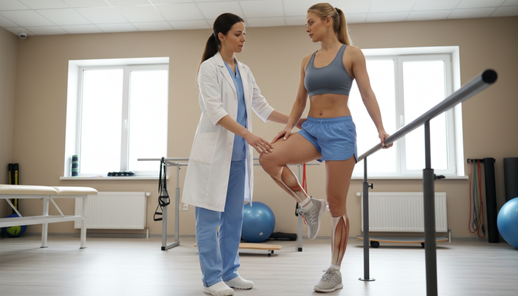
Hypermobility means a joint moves beyond the typical range expected for that joint. Joint laxity refers to looser-than-average ligaments or connective tissues that help restrain joint motion. These terms overlap, but they are not always identical.
A person can have:
Local hypermobility
One joint is unusually mobile, such as a shoulder that subluxates or a knee that hyperextends.
Generalized joint hypermobility
Multiple joints throughout the body move beyond the expected range.
Symptomatic hypermobility
Increased motion is associated with pain, instability, injury, fatigue, or functional limits.
A defined hypermobility syndrome
Hypermobility occurs as part of a broader connective tissue condition.
Many flexible people have no pain and never need orthopedic care. The problem begins when the joint’s passive restraints, such as the capsule and ligaments, do not provide enough control under load. Then the muscles must work harder to stabilize the joint. Over time, that can lead to pain, overload, inflammation, tendon irritation, cartilage stress, or injury.
In other words, joint laxity hypermobility is not automatically a disease. It becomes clinically important when it changes how the hip, knee, kneecap, pelvis, or lower limb functions.
Why connective tissue matters
Ligaments, capsules, tendons, labrum, meniscus, cartilage, and skin all contain connective tissue. In hypermobility, those tissues may stretch more easily or recover less predictably after strain.
For the hip and knee, this matters because these joints need both motion and stability.
The hip is a deep ball-and-socket joint. Its bony shape provides much of its stability, but the labrum, capsule, and surrounding muscles also play important roles. When the capsule is lax or the socket is shallow, the hip may experience microinstability.
The knee is more exposed. It depends heavily on ligaments, menisci, muscle control, limb alignment, and the tracking of the kneecap. In a hypermobile person, a small alignment issue may become more symptomatic because the soft tissues provide less restraint.
Hypermobility can be genetic, acquired, or activity-related
Some patients are born with generalized laxity. Others develop increased motion through years of flexibility training. Pregnancy, hormonal changes, prior injury, and certain medical conditions can also affect ligament behavior.
The clinical question is not simply, “Are the joints loose?” The better question is:
Is the extra motion contributing to pain, instability, tissue injury, or loss of function?
That distinction guides treatment.
Hypermobility syndromes and the Beighton score
Hypermobility exists on a spectrum. Some patients have benign flexibility with no medical concerns. Others have a heritable connective tissue disorder that affects more than the joints.
Common terms patients may hear include Ehlers-Danlos syndrome, Beighton, soft tissue laxity, loose joints, hypermobility spectrum disorder, and generalized joint hypermobility. The terminology can be confusing, especially because symptoms can overlap.
Ehlers-Danlos syndrome and related conditions
Ehlers-Danlos syndromes, often abbreviated EDS, are a group of heritable connective tissue disorders. Different types have different features and risks.
The type most often discussed in orthopedic and sports medicine settings is hypermobile Ehlers-Danlos syndrome, or hEDS. Patients with hEDS may have generalized joint hypermobility, pain, recurrent sprains, subluxations, fatigue, and soft tissue symptoms. Unlike some other EDS types, hEDS currently does not have a single widely used genetic test that confirms the diagnosis. Diagnosis is clinical and based on established criteria.
Other EDS types can involve more pronounced skin findings, arterial or organ risks, wound healing concerns, or other systemic features. Vascular EDS, for example, is a distinct and serious condition that requires specialized medical care.
Not every hypermobile person has EDS. Some patients fit better into the category of hypermobility spectrum disorder, often called HSD. HSD can still cause real orthopedic pain and functional issues, even if the patient does not meet criteria for hEDS.
The Beighton score
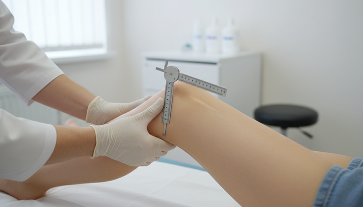
The Beighton score is a simple screening tool used to assess generalized joint hypermobility. It scores certain movements on a 9-point scale.
The test includes:
Beighton maneuver | Points |
Passive extension of the little finger beyond 90 degrees, right and left | 2 |
Passive thumb movement to the forearm, right and left | 2 |
Elbow hyperextension beyond 10 degrees, right and left | 2 |
Knee hyperextension beyond 10 degrees, right and left | 2 |
Forward bend with palms flat on the floor and knees straight | 1 |
Total possible score | 9 |
Common cutoffs vary by age. A score of 5 or more out of 9 is often used for many adults. Children and adolescents may require a higher cutoff because flexibility is more common at younger ages. Adults over age 50 may use a lower cutoff because mobility often decreases over time.
The Beighton score is useful, but it is not a complete diagnosis. It does not evaluate the shoulders, hips, feet, ankles, spine, or kneecaps in detail. It also does not measure pain, instability, tissue quality, or function.
A person with hip microinstability and a labral tear may have a modest Beighton score. Another person may score high and have no pain. That is why the orthopedic exam must focus on the painful joint, not just the total score.
Medical implications beyond the joints
When hypermobility is part of a syndrome, it may affect more than the musculoskeletal system. Depending on the individual condition, associated issues can include:
Skin that stretches more than expected or bruises easily
Delayed wound healing or wider scars
Dizziness or heart rate changes with standing
Gastrointestinal symptoms
Pelvic floor symptoms
Headache, neck pain, or fatigue
Medication sensitivity or anesthesia planning considerations
Some patients benefit from evaluation by rheumatology, genetics, cardiology, primary care, pain medicine, or other specialists. This does not mean every flexible person needs extensive testing. It means patients with widespread symptoms, concerning family history, unusual bruising, vascular concerns, or multi-system symptoms deserve a broader medical review.
For orthopedic planning, this background matters because connective tissue quality can influence injury risk, surgical decision-making, wound healing, rehabilitation pace, and pain recovery.
How hypermobility affects hip pain and hip preservation
The hip is often described as a stable joint because the femoral head sits inside the acetabulum, or socket. That is true when the bony socket, labrum, capsule, and muscles all work together. In a hypermobile patient, the soft tissue side of that system may provide less restraint.
This can create a pattern called hip microinstability. The hip may not fully dislocate. Instead, it may subtly translate or shift during activity. That small extra motion can irritate the labrum, capsule, cartilage, iliopsoas tendon, hip flexors, adductors, or muscles around the pelvis.
Common hip symptoms in hypermobility
Patients with hypermobility-related hip problems may describe:
Groin pain with pivoting, squatting, running, or getting in and out of a car
Clicking, catching, or locking
A sense of slipping, shifting, or giving way
Pain in the front of the hip after prolonged sitting
Lateral hip pain from muscle overload
Deep buttock or pelvic pain
Hip flexor tightness that keeps returning despite stretching
Fatigue with standing or walking
A common trap is to keep stretching a hip that already has too much motion. If the main issue is instability, repeated stretching may increase symptoms. Many patients do better with strength, control, and positional training rather than aggressive flexibility work.
Labral tears and hip microinstability
The labrum is a ring of fibrocartilage around the socket. It helps maintain the suction seal of the hip and contributes to stability. In a hypermobile hip, the labrum may experience repeated shear or traction forces.
This can contribute to labral tearing, especially when other structural factors are present, such as:
Hip dysplasia or borderline dysplasia
Femoroacetabular impingement, often called FAI
Increased femoral anteversion
Acetabular version abnormalities
Repetitive pivoting or high-flexibility sports
Prior trauma
A labral tear does not always require surgery. Many people have labral findings on MRI that are not the primary pain generator. The key is to match imaging with symptoms and exam findings.
For a hypermobile patient, the question is not only, “Is the labrum torn?” It is also, “Why did the labrum tear, and what is keeping the hip painful?”
If the labrum is compensating for instability, simply trimming torn tissue may not solve the underlying problem. Hip preservation care aims to protect the labrum, restore mechanics, and address instability when present.
Hip dysplasia and borderline dysplasia
Hip dysplasia means the socket does not cover the femoral head enough. This leaves the hip more dependent on the labrum and capsule for stability. In a patient with hypermobility, that combination can be especially important.
Symptoms of dysplasia can overlap with labral tears and hip flexor problems. Patients often describe groin pain, lateral hip fatigue, clicking, and pain with impact activity.
Plain x-rays are critical. MRI can show the labrum and cartilage, but x-rays show the bony coverage and alignment. When dysplasia is present, the main problem may be structural undercoverage rather than an isolated labral tear.
In more significant dysplasia, periacetabular osteotomy, commonly called PAO, may be considered. PAO reorients the socket to improve femoral head coverage while preserving the native hip joint. It is a major hip preservation surgery and is different from hip arthroscopy.
Some patients with borderline dysplasia and labral pathology may be candidates for arthroscopy with careful capsular management. Others need bony correction with PAO, sometimes with arthroscopy performed before, during, or after the bony procedure depending on the case. The decision depends on symptoms, imaging, instability signs, cartilage health, age, activity goals, and physical exam.
Non-surgical hip care
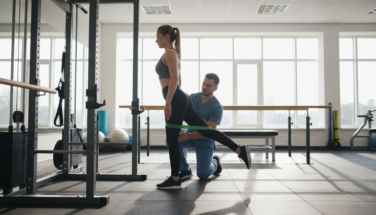
For many hypermobile hips, treatment begins with a focused rehabilitation plan. The goal is not to make the hip looser. The goal is to improve control.
A useful program often includes:
Gluteal and deep hip rotator strengthening
Core and pelvic control
Single-leg balance and movement retraining
Avoidance of repeated end-range stretching
Gradual return to running, dance, lifting, or sport
Education on positions that provoke instability
Treatment of tendon overload around the hip
Physical therapy should be specific. General stretching, generic strengthening, or repeated manipulation may not address microinstability. Progress often takes time because the muscles are being asked to provide stability that the passive tissues do not fully provide.
How hypermobility affects knee pain and knee injuries
The knee has less bony constraint than the hip. It depends on the ACL, PCL, MCL, LCL, menisci, capsule, tendons, muscles, and limb alignment. Hypermobile soft tissues can change how the knee handles cutting, landing, stairs, squatting, and prolonged standing.
The most common knee issues I see in hypermobile patients involve kneecap tracking, knee hyperextension, recurrent sprains, and overload pain.
Patellar instability and kneecap pain
The patella, or kneecap, must glide in a groove at the end of the femur. If the soft tissues are lax, the kneecap may shift more than expected. Some patients have true dislocations. Others feel the kneecap slide or catch without a full dislocation.
Risk factors can stack together:
Generalized ligamentous laxity
Shallow trochlear groove
Patella alta, where the kneecap sits high
Increased femoral anteversion
Increased tibial tubercle distance
Flat feet or rotational alignment differences
Hip weakness or poor landing mechanics
Symptoms may include anterior knee pain, swelling after activity, apprehension with pivoting, or a sense that the kneecap might “go out.”
Rehabilitation focuses on hip strength, quadriceps control, foot and ankle mechanics, and movement retraining. Bracing or taping can help some patients, especially during higher-risk activities.
When instability is recurrent, surgical options may be considered. Procedures can include medial patellofemoral ligament reconstruction, tibial tubercle osteotomy, or trochlear procedures in selected cases. In hypermobile patients, the surgeon must account for tissue quality, alignment, and the risk of recurrent laxity. Soft tissue reconstruction alone may not be enough if major bony risk factors are present.
Knee hyperextension and overload
Many hypermobile patients stand with their knees locked back. This posture may feel easy because it uses passive ligaments rather than active muscle control. Over time, it can irritate the back of the knee, overload the capsule, strain the hamstrings or calf, and affect kneecap mechanics.
In sports, landing into knee hyperextension can increase stress on the ACL and posterior capsule. During daily life, constant locking may contribute to fatigue and pain.
Treatment often includes learning a “soft knee” stance, strengthening the quadriceps and hamstrings, and improving hip control. Some patients benefit from temporary bracing or proprioceptive training so they can feel where neutral knee position is.
Meniscus, ligament, and tendon problems
Hypermobility does not guarantee a ligament tear, but it can influence injury patterns. If the knee moves farther before the muscles react, tissues may experience higher strain during a twist, fall, or landing.
Possible knee problems include:
Recurrent MCL or ankle sprain patterns that affect knee mechanics
ACL injury risk in pivoting sports
Meniscus irritation or tearing after twisting episodes
Pes anserine, patellar tendon, or quadriceps tendon overload
Iliotibial band irritation from poor limb control
Swelling after activity because the joint is repeatedly irritated
The best care plan looks at the entire limb. A painful knee may reflect hip weakness, foot mechanics, rotational alignment, or a movement pattern that places the knee at the end of its available range.
For both hip and knee conditions, the goal is joint preservation. That means reducing unnecessary stress, correcting structural problems when needed, protecting cartilage and labral or meniscal tissue, and keeping the patient active in a sustainable way.
Work-up and surgical planning for hypermobile hip and knee patients
A thoughtful work-up begins with listening. Patients with hypermobility often have a long history of being told their imaging is “not that bad” or that flexibility is always beneficial. The exam should connect symptoms, movement, imaging, and tissue behavior.
History and physical exam
Key history points include:
Childhood flexibility, dance, gymnastics, martial arts, cheer, or yoga background
Prior dislocations, subluxations, sprains, or “party tricks”
Family history of hypermobility, aneurysm, sudden vascular events, or diagnosed connective tissue disorders
Skin findings, bruising, delayed healing, dizziness, fatigue, or widespread pain
Hip clicking, instability, locking, or groin pain
Kneecap instability, knee hyperextension, swelling, or giving way
Response to prior physical therapy, injections, braces, or surgery
The physical exam usually includes Beighton scoring, gait assessment, limb alignment, hip range of motion, impingement testing, instability testing, strength testing, kneecap tracking, knee ligament exam, and functional movements such as squatting or single-leg stance.
No single test tells the whole story. A positive impingement test may coexist with instability. A labral tear may coexist with dysplasia. A kneecap tracking problem may reflect hip rotation and generalized laxity, not just the knee itself.
Imaging and medical evaluation
Orthopedic imaging depends on the painful joint.
For the hip, evaluation may include:
Standing pelvis and hip x-rays
Specialized views to assess cam or pincer morphology
Measurements of acetabular coverage and version
MRI or MR arthrogram to assess the labrum, cartilage, capsule, and surrounding soft tissues
CT scan in selected cases to study femoral version or complex bony anatomy
For the knee, evaluation may include:
Standing alignment x-rays
Patellar tracking views
MRI for cartilage, meniscus, ligament, or MPFL injury
Rotational or advanced imaging in selected instability cases
Lab testing is not required for every patient with hypermobility. It may be appropriate when symptoms suggest inflammatory arthritis, autoimmune disease, infection, metabolic disease, or another non-mechanical source of pain. Rheumatology evaluation can help when there is widespread pain, swelling in multiple joints, morning stiffness, systemic symptoms, or uncertainty about diagnosis.
Genetics or cardiology evaluation may be appropriate when the history suggests a heritable connective tissue disorder with vascular or cardiac implications. For some patients with suspected EDS or related conditions, documenting the diagnosis helps guide anesthesia, wound care, rehabilitation, and long-term medical monitoring.
Hip arthroscopy in hypermobile patients
Hip arthroscopy can be helpful for selected patients with labral tears, FAI, loose bodies, cartilage flaps, or certain instability patterns. In hypermobile patients, the details matter.
The hip capsule is a key stabilizer. During arthroscopy, the surgeon often makes a capsulotomy to access the joint. In a patient with soft tissue laxity, leaving the capsule open or too loose may contribute to postoperative instability or persistent pain.
Specific measures may include:
Labral repair rather than simple removal when tissue quality allows
Preserving or restoring the labral seal is especially important when stability is a concern.
Capsular closure or capsular plication
The capsule may be repaired and tightened to improve stability. Plication is often considered when laxity or microinstability is present.
Careful correction of FAI
Cam or pincer bone can damage the labrum, but over-resection can create new problems. The goal is balanced motion without instability.
Protection of the capsule during surgery
Surgical planning may use smaller or more controlled capsulotomies when appropriate.
Postoperative bracing
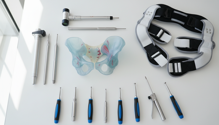
A brace may limit extension, external rotation, or other positions that stress the capsule during early healing.
Modified rehabilitation
Physical therapy may progress more cautiously. Early rehab often protects the repair, avoids aggressive stretching, and emphasizes controlled strengthening.
The surgical plan should match the diagnosis. Arthroscopy for a labral tear in a stable hip is different from arthroscopy for a hypermobile hip with microinstability. It is also different from arthroscopy in the setting of frank dysplasia.
When bony correction is needed
Soft tissue procedures cannot always solve a bony stability problem. If the socket is too shallow, as in hip dysplasia, the labrum and capsule may be overloaded because they are compensating for inadequate coverage.
In that setting, repairing the labrum without correcting the undercoverage may leave the main mechanical problem untreated. For appropriate candidates, periacetabular osteotomy (PAO) can improve coverage and reduce overload on the labrum and cartilage. This is one of the most important distinctions in hip preservation.
The same principle applies around the knee. Recurrent kneecap instability may not respond well to isolated soft tissue tightening if the underlying bony alignment is a major driver. Procedures that address tibial tubercle position, patella height, or trochlear anatomy may be considered in selected patients.
Setting expectations after surgery
Hypermobility does not mean surgery cannot work. It does mean the margin for error may be smaller. Accurate diagnosis, careful tissue handling, stable repair, and a tailored rehabilitation plan become more important.
Recovery may also require patience. Hypermobile patients can be strong and athletic, but their tissues may respond differently to load. Some need slower progression, longer bracing, closer physical therapy guidance, or more attention to pain regulation and fatigue.
The goal is not to make the joint stiff. The goal is to restore enough stability so the joint can move with less pain and less injury risk.
The practical takeaway for painful loose joints
Hypermobility changes the way hip and knee problems should be evaluated. A labral tear, kneecap instability, or knee pain pattern may look familiar on the surface, but the treatment plan should account for soft tissue laxity, bony anatomy, muscle control, and any broader medical features.
The most useful approach is stepwise:
Confirm whether hypermobility is local or generalized.
Identify whether pain comes from instability, impingement, dysplasia, tendon overload, cartilage injury, or another source.
Use x-rays and MRI thoughtfully, not in isolation.
Start with targeted stability-based rehabilitation when appropriate.
Consider medical evaluation when symptoms suggest a connective tissue syndrome or inflammatory condition.
If surgery is needed, plan around tissue quality, capsular stability, labral or meniscal preservation, bracing, and any needed bony correction.
For patients with hip or knee pain and hypermobile joints, the main message is reassuring: flexibility does not have to mean fragility. With the right diagnosis and a plan that respects joint stability, many people can reduce pain, prevent repeat injury, and stay active while protecting the hip and knee for the long term. A specialist like world-renowned hip and knee specialist Atul Kamath, MD can help with the triage, management, and diagnosis of hypermobile joint symptoms.


Comments